Anal Sacculectomy
The anal sacs are pockets situated either side of the anus which vary in size, depending on the size of the dog. They have small openings towards the opening of the anus. Their purpose is to hold secretions from glands and are occasionally known as “scent glands”, as the liquid they hold is a very potent and offensive smell.
These sacs tend to empty their contents when a dog passes faeces. On occasion, they can spontaneously empty when a dog barks or jumps. Or can even do so without any sudden activity.
Problems with Anal Sacs
The anal sacs can become problematic for several reasons:
Page 4 - Anal Sacs
-
Impaction: This is Where the Sacs Fail to Empty Naturally, so the Liquid Secretions Inside Build-Up, Causing the Dog to Feel Discomfort. The Dog May Lick at its Rear End or Drag their Bottom Across the Floor to Release the Pressure
-
Anal Sac Infection
-
Anal Sac Rupture: If Not Emptied, the Gland May Eventually Rupture, Causing the Foul Liquid to Run into the Tissues Around the Bottom, Causing a Painful Infection and Abscessation. The Rear End Will Be Extremely Hot, Swollen, and Painful. The Abscess May Eventually Burst, causing a Hole to Appear in the Skin Near the Bottom. Before the Abscess Bursts, the Infection Can Make the Patient Extremely Unwell.
-
Anal Sac Adenocarcinoma: Cancer of the Anal Sac
Anal Sac Impaction
Many dogs experience anal sac impaction, they may go on to express the impacted material spontaneously or after rubbing the area. If the pressure does not relieve; manual expression may be required, whereby the liquid inside the sac is squeezed out. If the material is not easily expressed, the vet may recommend flushing of the gland. Sometimes a diet change may be recommended to help the problem.
For cases that suffer from frequent impactions or multiple infections or rupture, the vet may recommend that the glands are flushed and filled with a mixture of anti-inflammatory and antibiotic. In more severe cases or cases which involve cancer of the sac, a surgical cure is to perform an Anal Sacculectomy. The surgery involves complete removal of the anal sacs.
Pre-Operative Considerations:
Page 4 - Pre-Operative Considerations:
-
Patients Must Be of Good General Health and Able to Undergo a General Anaesthetic
-
Scar Tissue is Likely to Make the Surgery More Challenging and Can Increase the Likelihood of Short-Term Problems After the Operation
-
We Ask That All Patients Are Starved for 24 Hours Before the Operation and Taken Out for Toilet Purposes Before Attending the Practice on the Day of the Operation
-
If There Has Been Anal Sac Rupture or Abscessation, the Surgery Cannot Go Ahead Until the Area is Fully Healed
The Surgical Procedure
The patient undergoes a pre-operative examination, then a combination of sedative/painkiller is administered. The patient is then anaesthetised, and the rear end is clipped short and cleaned. The anal sacs are flushed with antiseptic. The vet prepares a resin which is instilled into the sacs, making them easier to surgically dissect. A short incision is made into the skin over the gland, and the surgeon gently dissects the sacs out from between the muscle layers around the rectum. Once the sac is removed, sutures are placed to close the space, these may either be dissolving sutures underneath the skin, or skin sutures which sit on top of the skin. The patient is cleaned and recovered from anaesthesia by the nursing team.
What to Expect Immediately After Surgery
Any surgery around the rectum and the rear end is very uncomfortable. We supply pain relief by injection, while the dog is in the surgery, and often an Elizabethan Collar is required. Preventing the dog from licking at the area and worsening the discomfort and soreness. Pain relief will be provided for use at home, however, in some cases, antibiotics may be required.
The recovery phase differs, tremendously, for this procedure. Some dogs act as if nothing has happened and return to normal within 24 hours of the procedure, whereas some refuse to pass faeces for several days post-operation. Other patients exhibit diarrhoea after the surgery, often when scarring is present, and the tissues have had to be manipulated extensively. If this is the case, then you may be required to keep the dog restricted to a small area and clean the rear end regularly. This will resolve in a matter of days, and medication can be provided to help reduce the volume of diarrhoea. Some dogs lose some ability to hold faeces and may need to poo more regularly. We recommend frequent opportunities for toileting in the week post-operation, or for the dog to be kept restricted to an area, where the furnishings can be more easily cleaned. For a few weeks post-surgery, your dog may have accidents, where faeces are passed unexpectedly, this usually occurs when they bark or jump up and is caused by the swelling and tissue disruption during the surgery. In majority cases, this resolves over a few weeks. Very occasionally, we see infections in the surgical sites, this is made more likely if the patient can lick and interfere with the site and should be treated with bathing and antibiotics.
Long Term Expectations
We expect a full recovery within a month of the procedure and return to normal toileting patterns. With any surgery in this area, there is a risk of long-term incontinence. This is a rare long-term side effect, and usually where a very large dissection has been required to remove a cancer of the anal sac. If the anal sac has ruptured before surgery, there may be small pieces of the sac tissue within the surgical site that aren’t removed, and these may then cause an abscess or irritation after the surgical site has healed, this would mean a second surgery was needed. This only happens very rarely.


Ear Canal Ablation
Sometimes an ear infection is so severe that surgery is the only option. The organism growing may resist treatment, the ear canal might have mineralized due to chronic irritation, or the ear canal might be so scarred and narrowed that external cleaning is impossible. No matter the issue, this degree of an irreversible disease requires surgical treatment. In such cases, all the diseased tissue, the entire ear canal, bones of the middle ear, etc. are removed. The middle ear is drained, and the healthy tissue around the ear is closed. This ends what has generally been a long ordeal of pain, odour, ear cleaning, and expensive veterinary medications, and re-checks.
It is important to understand the surgical procedure and its associated risks. This surgery requires advanced skill and is a procedure that not all veterinarians are comfortable performing. The surgery essentially removes the ear structures but leaves the ear flap unchanged. The round bone behind the ear called the tympanic bulla is opened and flushed. A normal bulla is hollow and air-filled. After years of otitis, the bulla is usually packed with pus, slime, or infectious material that must be scooped or rinsed out. Many important nerves travel through this area of the ear, and these are exposed to potential damage during surgery.
Preparing for the TECA
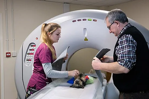
Radiographs or a CT scan to assess the tympanic bullae are helpful. It is useful to know before surgery how bad the bullae look, how narrowed the ear canals are and if they are mineralized, and if there is an obvious tumour growing. These images help confirm that the surgery is appropriate for the patient.
The ear may be cultured, and this helps get the patient on an effective antibiotic right from the beginning. Further cultures may still be required once the bullae are opened. It is important to assess the patient's cranial nerve function before surgery. If these nerves are diseased before surgery, it is unlikely that they will regain function after surgery. Nerve disease that results from surgery is usually temporary, so it is important to know if the nerve problems exist before surgery.
The facial nerve runs just near the base of the ear, and this nerve controls facial expression. Facial paralysis is not uncommon after long-standing ear disease. This means that the patient is slack-jawed, usually on one side of the face, and may not be able to blink. After a time, the eye usually retracts into the eye socket to facilitate tear lubrication, so that the loss of blinking does not lead to eye damage. Initially, lubricating gels are helpful.
Hearing is usually diminished after long-term ear infections. Further hearing loss after ear ablation may not represent a dramatic change for the patient. Most owners have a good sense of whether their pet can hear, so it is rarely necessary to formally test hearing. After the ear canals are ablated, in many patients, some hearing remains as sound waves can still be transmitted through the tissues.
What Happens in Surgery
Before surgery, the ears and head are shaved, and the ear canal is flushed one last time to remove as much infected material as possible. This is done to minimize bacterial contamination of the normal tissue.
Both the vertical and horizontal portions of the ear canal are removed as one long intact curved cylinder. The bones of the middle ear and the eardrum are removed as well, with the bone of the tympanic bulla exposed and opened. Any material is flushed out, and the cellular lining of the bone is scraped away. Any material left inside after closure will lead to chronic drainage of liquid from the incision. Sometimes an external drain is left in place during the healing period.
Oral antibiotics and pain medication can be expected after surgery, as can an Elizabethan collar to protect the delicate incisions from scratching.
Cat-Friendly Clinic
We are proud to announce that Willow Veterinary Clinic Ltd has recently received Bronze Cat-Friendly Clinic status at our Endon branch. This is a scheme run by the International Society of Feline Medicine (the veterinary division of International Cat Care), to help veterinary practices cater to the unique needs of cats. We aim to provide an excellent standard of care to all our feline patients, whilst also adhering to the principles of the scheme.
One of the main aims of the scheme is to reduce any stress that cats may experience during a visit to the vets. We vow to handle cats with gentleness and respect, to minimise fear and anxiety during handing, examination, and procedures. We also ensure that cats that need to stay with us in the clinic are hospitalised in a cat-friendly environment.
Plus, we can offer cat-only appointments for nervous cats. If you would like any further information, please feel free to contact the surgery and speak to our Cat-Friendly Advocate, Janet Carroll. Alternatively, please visit www.icatcare.org to find out more.
Janet is our Cat Advocate for the ISFM Cat-Friendly Clinic scheme, ensuring that all cat's needs are met by the practice. We are proud to have a nurse with the ISFM DipFN qualification in our nursing team, and we're sure that our feline patients will appreciate her skills and expertise.
Kidney Disease in Cats
Did You Know that Kidney Disease Affects up to 50% of Cats Over the Age of 15 Years?
Although kidney disease also affects dogs, it is seen about three times more frequently in cats. Unfortunately, this is a progressive disease and clinical signs only appear once the kidneys have already lost most of the normal function.
Signs can include:
Page 3 - Kidney Disease
-
Weight Loss
-
Poor Appetite
-
Lethargy
-
Increased Thirst
-
Increased Urination
-
Poor Coat Condition
-
Vomiting
-
Halitosis (Bad Breath)
Although there is no cure, early diagnosis enables supportive treatment to be administered, which can help to arrest the progression of the disease, giving your pet a better quality of life.
Steer Clear of Human Medicines
Did you know that just one 500mg paracetamol tablet is enough to be toxic and fatal to a cat? This includes flu capsules that contain paracetamol.
Occasionally, a curious cat will play with and chew a tablet or capsule that hasn't been stored out of reach. On other occasions, it has been a well-meaning owner that has given the tablet. Cats are very sensitive to paracetamol, much more so than humans and dogs. When the drug is broken down by the liver in a cat, a toxic chemical is produced which results in several complications.
These include severe liver damage and a reduction in the ability of red blood cells to carry oxygen around the body.
Signs of paracetamol toxicity include:
Page 3 - Steer Clear of Human Medicines
-
Gums Becoming a Bluish Colour
-
A Very Fast Heart Rate
-
Becoming Very Quiet and Depressed
-
Difficulty in Breathing
-
Vomiting
-
Passing Dark Brown Urine
-
Skin May Start to Look Yellow
-
Swelling of the Paws and Face
Prognosis depends on how quickly the cat is taken to the vets and how much paracetamol has been ingested. Unfortunately, due to the toxic effects on cats, it is highly possible they may not survive.
Bringing Your Cat to the Practice
By nature, cats are independent, territorial animals that need to be in control of their surroundings. They are sensitive to different smells, sounds, and sights. All these different senses can make bringing your feline friend to the practice a very stressful experience.
Tips to help reduce stress can include:
Page 3 - Bringing Your Cat to the Practice
-
Making Sure You Use a Good Pet Carrier; Top Opening Ones Are Easier as the Cat Can Be Lifted Out Gently
-
Familiar Smells Can Help, Keep the Carrier Out at Home, so the Cat Can Use the Carrier as a Bed and Feel at Ease Around it
-
Don't Introduce the Carrier Only When There is a Visit to the Vet
-
Place Bedding or Clothing That Smells of Home into the Carrier
-
Spray the Carrier with Feliway, a Pheromone Calming Spray, 30 Mins Before Using
-
If Your Cat Becomes Very Stressed, Try Wrapping Them in a Thick Towel Before Placing Them into the Carrier
When travelling, avoid loud noises such as the radio. Cover the carrier with a blanket or towel during the journey to make them feel calmer. Secure the carrier in the footwell, so that it isn’t likely to move, or hold the carrier carefully and securely to avoid swinging. Finally, take your cat to a veterinary practice that has been recognised as Cat-Friendly, like Willow Veterinary Clinic Ltd.
Arthritis in Cats
Until relatively recently, arthritis in cats was not commonly recognised or treated. Cats are masters at disguising pain, due to their natural survival instinct. However, a recent study showed that 90% of cats, over 12 years of age, had some form of arthritis.
How to tell if your cat is affected:
Page 3 - Arthritis in Cats
-
Hesitant When Jumping Up or Down
-
Jumping from Lower Heights
-
Difficulty with Going Up or Downstairs
-
Stiffness, Especially After Sleeping or Obvious Lameness
-
Difficulty Using the Litter Tray
-
Difficulty in Getting Through the Cat Flap
-
Reduced Activity
-
Sleeping in Different Places, Usually Easier to Access
-
Increased Grooming Over Specific Places, or Inability to Groom
-
Irritable and Grumpy When Stroked or Picked Up
If you suspect your cat could be arthritic, they must have a full check by a veterinary surgeon, so that treatment and medication can begin.
To help at home:
Page 3 - Arthritis in Cats (B)
-
Place Food and Water in Easily Accessible Places
-
Provide Easily Accessed Comfortable, Warm Beds
-
Ensure Cat Flaps Are Easy to Open
-
Use Lower Sided Cat Litter Trays for Easier Access
-
Spend Time Grooming Them If It Has Become Difficult for Them
-
Regular Clipping of Any Overgrowing Nails
Antifreeze
As the weather grows colder in the winter months, antifreeze poisoning in cats becomes more common. The toxic substance is ethylene glycol and is found in products such as screen washes and de-icers. Cats are attracted to the 'sweet' taste in this product, and even a small amount can cause severe kidney damage and be fatal.
Clinical signs you may see, include:
Page 3 - Antifreeze
-
Vomiting
-
Unsteady on Feet
-
Weakness
-
Excessive Drinking
-
Excessive Urination or No Urination
-
Collapse
If not treated promptly, this can be fatal. If you think your cat could have ingested antifreeze, contact your vet as soon as possible.
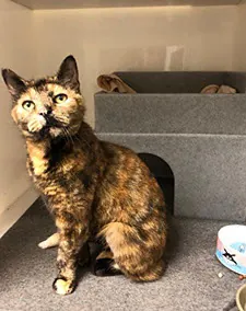
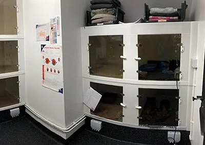
Introducing Our New Cat-Friendly Ward
We have recently finished our brand-new cat ward in our Endon practice. This is a room that has been designed following the ISFM feline-friendly guidelines, to provide the very best accommodation and stress-free environment for our feline patients.
We have spacious kennels, allowing plenty of room to keep the sleeping, litter, and food/water areas separate. For long-stay patients, we can open a ‘cat flap’ between the kennels, providing double the amount of space. The fronts are made with tinted glass, allowing the patients to watch the goings-on without looking through bars. With the rest of the kennel made from a specialised plastic, this keeps them warmer and without any reflections, which can cause stress.
We have also recently purchased some ‘cat hides’ that allow our patients to hide under them or sit on top, helping them to feel secure and safe.
Not only is this room used for accommodation, but we also take blood samples, perform full clinical examinations, take blood pressures, and take temperatures solely in this room. This also helps our feline patients stay calm and relaxed, as they are kept in one location instead of being moved from room to room.
We understand the importance of providing the best possible care, and we’re always striving to improve our facilities to achieve this.
For help or advice with any feline problems, we provide feline nurse clinics with our cat-friendly nurse, Janet. Please ring the surgery to book an appointment.